
Figure 2. Steric-zipper protofilaments.
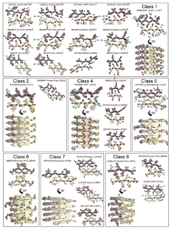
Twenty-eight atomic structures of steric-zipper protofilaments from amyloid-forming proteins, determined by X-ray diffraction. All are viewed projected down the protofilament axis, revealing the two sheets (one gold and one purple) with their interdigitated sidechains. Selected zippers are also viewed perpendicular to the protofilament axis, with five layers of β-strands shown with backbones as arrows. Water molecules are shown as aqua spheres; notice their absence from the interfaces between the paired sheets.