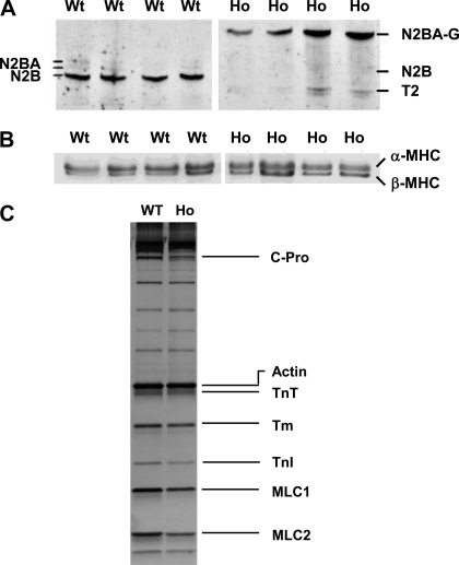
Fig. 2.
SDS-PAGE analysis of wild-type (WT) and homozygote (Ho) rat ventricles. Typical expression profile of titin isoforms (A), myosin heavy chain isoforms (B), and other myofilament proteins (C) in WT and Ho ventricles examined by SDS electrophoresis as described in methods and materials. A: left ventricles (4 each) from WT and Ho rats were analyzed for titin isoforms. WT ventricles expressed predominantly a shorter N2B isoform whereas the Ho ventricles expressed predominantly a giant N2BA-G isoform. T2 is the degradation product of titin. B: WT ventricles expressed 68 ± 4% α- and 32 ± 4% β-myosin heavy chain (MHC) and the Ho ventricles expressed 59 ± 4% α- and 41 ± 4% β-MHC. C: no major differences were visible in expression profile of other myofibrillar proteins between WT and Ho ventricles. C-Pro, myosin binding protein-C; TnT, cardiac troponin T; α-Tm, α-tropomyosin; TnI, cardiac troponin I; MLC-1, ventricular myosin light chain 1; MLC-2, ventricular myosin light chain 2. Protein loading was not homogenous between lanes.

