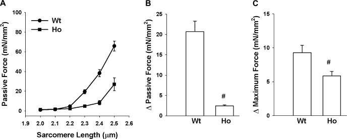
Fig. 4.
Magnitude of sarcomere length (SL)-dependent changes in passive and maximum Ca2+-activated force in WT and Ho trabeculae. A: passive force was measured by sequentially stretching WT (n = 4; ●) and Ho (n = 4; ■) trabeculae to various SL in pCa 9.0 solution. B and C: passive and maximum Ca2+-activated forces were measured in pCa 9.0 and 4.5 solution respectively, first at SL 2.0 μm and then at 2.35 μm in WT (n = 13) and Ho (n = 11) trabeculae. Magnitude of SL-dependent changes in passive force (B) and SL-dependent changes in maximum Ca2+-activated force (C) was determined by subtracting values recorded at SL 2.0 μm from those recorded at 2.35 μm. Open bar represents the mean and the error bars the SE. #Significantly different from values determined in WT trabeculae.

