Fig. 1.
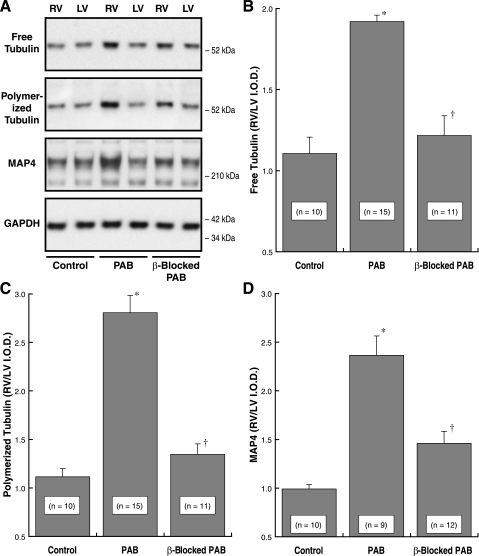
The effects of β1- and β2-adrenergic receptor blockade on components of the cardiac microtubule network during pressure overload hypertrophy. A: example immunoblots from the right ventricles (RVs) and left ventricles (LVs) of a pair of cats with 4 wk of pulmonary artery band (PAB)-induced RV hypertrophy; 1 animal was β-blocked with propranolol for the final 2 wk, and the other was not β-blocked. A monoclonal anti-β-tubulin antibody (clone DM-1B; Abcam) was used for the tubulin blots, our anti-myocardial microtubule-associated protein (MAP)4 antibody (43) was used for the MAP4 blot, and a monoclonal anti-GAPDH antibody (clone 6C5; Upstate Biotech) was used for the loading control blot. B–D: summary data for the background-corrected integrated optical density (I.O.D.) ratio of RV to LV in immunoblots from the indicated numbers of normal control cats, PAB cats without β-blockade, and PAB cats with β-blockade. *P < 0.05 for difference from control; †P < 0.05 for difference from PAB by 1-way ANOVA with Bonferroni post hoc analysis. For the LVs alone, there was no within-group difference for any of these 3 variables by 1-way ANOVA with Bonferroni post hoc analysis (P = 0.69 for LV free tubulin; P = 0.43 for LV polymerized tubulin; P = 0.29 for LV MAP4; each P value is the minimum value for the 3 comparisons within that group).

