Fig. 1.
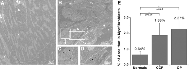
Electron microscopy of myofibroblasts in infarcted hearts. Normal myocardium (A) has almost no identifiable myofibroblasts, but following 5 days of coronary occlusion (B) myofibroblasts are readily identifiable by their large nuclei (N) as well as by their extensive endoplasmic reticulum (inset C) and the presence of stress fibers (inset D). Quantification of myofibroblasts (E) in different regions of the re-entrant circuits [common pathway (CCP) and outer pathway (OP)] showed that both regions had significant increases in the levels of myofibroblasts when compared with normal (0.64% in normal, 1.88% in CCP, and 2.27% in OP), but there was no difference in myofibroblasts levels in the CCP when compared with the OP, indicating that the electrical pathway does not affect myofibroblasts localization (n = 5).

