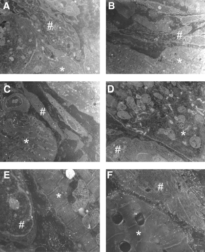
Fig. 3.
Electron micrographs of 5-day infarcted epicardial border zone (EBZ) with myocytes (*) and myofibroblasts (#) (A–F). Several examples of myocytes next of myofibroblasts showed no gap junctional plaques between these 2 cell types, even at the longitudinal ends of the myocytes (for example, see C).

