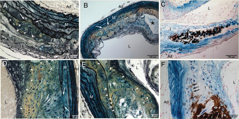
Figure 1.
Development of cartilaginous metaplasia and calcification in atherosclerotic vessels of LDLr−/− mice. Aortic arches were dissected from SM22α-Cre+/0:R26R+/0:LDLr−/− mice fed with HFD for 18–20 weeks (A–C) and 24 weeks (D–F). Cells of SM origin were stained by X-gal before embedding. Calcification was stained by the von Kossa method (C and F). Cells of chondrocyte morphology were visualized by Movat pentachrome staining (A, B, D, and E). White arrows designate elastic lamina breakage. White arrowheads designate chondrocyte-like cells. Black arrows designate medial calcification. Asterisk designates necrotic core. L, lumen; I, intima; M, media; Ad, adventitia.

