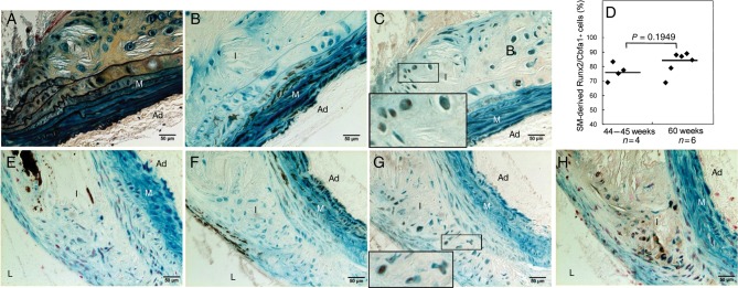
Figure 5.
SMCs gave rise to osteochondrogenic precursor- and chondrocyte-like cells in atherosclerotic ApoE−/− vessels. Aortic arches were dissected from 45-week-old (A–C) and 60-week-old (E–H) SM22α-Cre+/0:R26R+/0:ApoE−/− mice. Cells of SMC origin were stained by X-gal before embedding. Adjacent sections were stained by Movat pentachrome (A), von Kossa (E), and immunohistochemistry for SM22α (B and F), Runx2/Cbfa1 (C), Sox9 (G), and Col II (H). Insert in C and G. Higher-powered magnification of the boxed region shows colocalization of β-galactosidase (blue) with osteochondrogenic marker Runx2/Cbfa1 (C, brown) or with chondrocytic transcription factor, Sox9 (G, brown). (D). The percentage of Runx2/Cbfa1-positive cells in calcified atherosclerotic vessels of LDLr−/− mice. L, lumen; I, intima; M, media; Ad, adventitia.

