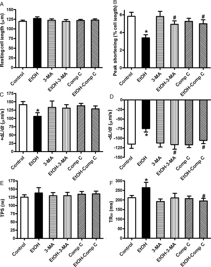
Figure 5.
The effect of 3-MA and compound C on ethanol-induced cardiomyocyte contractile defects. Freshly isolated cardiomyocytes from WT mice were incubated with ethanol (240 mg/dL) in the presence or absence of 3-MA (10 mM) or compound C (5 µM) prior to mechanical assessment. (A) Resting cell length; (B) peak shortening (normalized to the resting cell length); (C) maximal velocity of shortening (+dL/dt); (D) maximal velocity of re-lengthening (−dL/dt); (E) time-to-peak shortening (TPS); and (F) time-to-90% re-lengthening (TR90). Mean ± SEM, n = 55–70 cells from three mice per group, *P < 0.05 vs. control group, #P < 0.05 vs. ethanol (EtOH) group.

