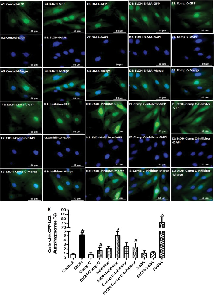
Figure 6.
The effect of 3-MA, compound C and mixed lysosomal inhibitor on ethanol exposure-induced autophagosome formation and autophagic flux in H9c2 myoblasts. H9c2 cells were transfected with adenovirus for 24 h to express the GFP-LC3 fusion protein. Cells were then exposed to ethanol (240 mg/dL) for 4 h in the absence or presence of the autophagosome inhibitor 3-MA (10 mM), the AMPK inhibitor compound C (5 μM), or lysosomal inhibitors. Rapamycin (5 μM) served as the positive control. DAPI staining was used for identification of the nucleus. Representative images of GFP (A1–J1), DAPI (A2–J2), and the merged images (A3–J3) depicting GFP-LC3 puncta in H9c2 cells; and (K) percentage of cells with autophagosomes. Cells with 10 or more punctate spots were scored as positive for autophagosomes. Mean ± SEM, n = 300–400 cells per group, *P < 0.05 vs. control group, #P < 0.05 vs. ethanol (EtOH) group.

