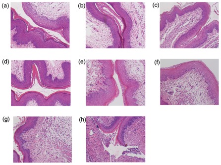
Figure 4. Histopathological examination of vaginal tissues after intravaginal application of SFT gel and other microbicide candidates.
Slides were prepared from vaginal tissues treated with placebo (1.5% HEC gel) (a), 0.03 mM SFT gel (b), 0.3 mM SFT gel (c), 3 mM SFT gel (d), 1% TFV gel (e), 3% carrageenan gel (f), 6% CS gel-treated group (g), or 1% N-9 gel-treated group (h). Hematoxylin-eosin staining was used for all slides. Magnification, ×100.

