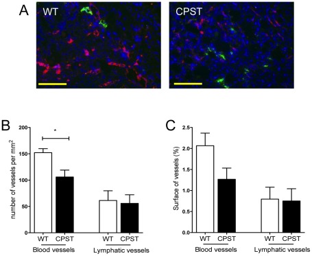
Figure 3. Impaired angiogenesis in CPST wounds.
Wound sections (day 7) of CPST and WT mice were double-labeled for CD31 (red) and Lyve1 (green). Nuclei were stained with DAPI (blue). Representative photos of wound granulation tissue in WT and CPST mice are depicted in (A). Number (B) and surface (C) of blood and lymphatic vessels in the wound beds. n = 7 per group. Scale bar (A) 100 µm. Mann-Whitney test, *P<0.05. (B and C) mean +/−SEM.

