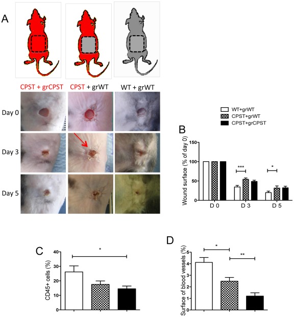
Figure 4. Impaired cell recruitment in the wound beds of CPST mice.
WT Skin flaps were transplanted on the back of CPST recipients (CPST+grWT) with corresponding controls (a). 2 months after transplantation, excisional biopsies were performed on grafts and monitored over time. Photographs (A) and surface (B) of the wounds at different time points. Number of CD45 positive cells (C) and surface of CD31+ blood vessels (D) were evaluated. n = 6 per group. Mann-Whitney test, *P<0.05; **P<0.01. (B–D) mean +/−SEM.

