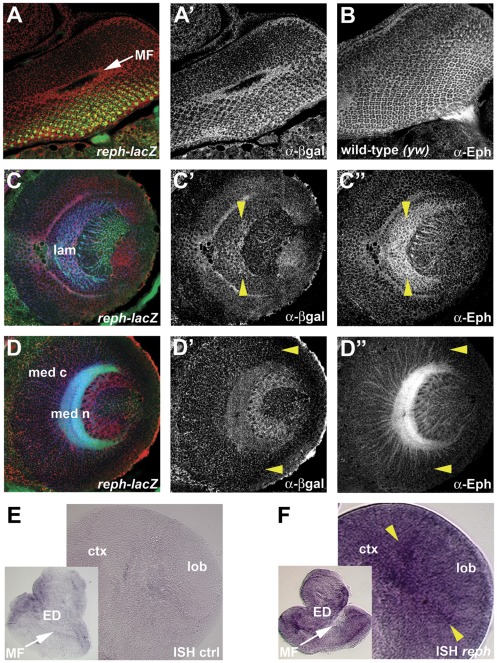
Figure 6. reph expression correlates with Eph expression in the developing optic lobe.
Expression patterns for reph in the developing retina, lamina and medulla were determined using a lac-Z reporter associated with rephk8617A and by in situ hybridization. Staining for HRP (anti-HRP, green, A,C,D) was used to visualize general cellular architecture. In the developing retina, reph (anti-β-gal, red in A, shown alone in A′) was expressed primarily in cells within and posterior to the MF, which correlated well with Eph expression (anti-Eph, B). In the lamina, reph was expressed in a high-midline, low-dorsoventral gradient (C′, demarcated by yellow arrowheads), as was the case for Eph (C″). In the medulla, reph expression within cortical neurons diminished at the dorsoventral margins (D′, yellow arrowheads) as was the case for Eph expression (D″). In situ hybridization (controls are shown in E) supported the lacZ reporter data. In the developing retina reph expression was found in cells within and posterior to the MF, while within the lamina region reph expression was highest at the midline (F, yellow arrowheads). Abbreviations: cortex (ctx), eye disc (ED), lamina (lam), lobula (lob), medulla cortex (med c), medulla neuropil (med n), morphogenetic furrow (MF).

