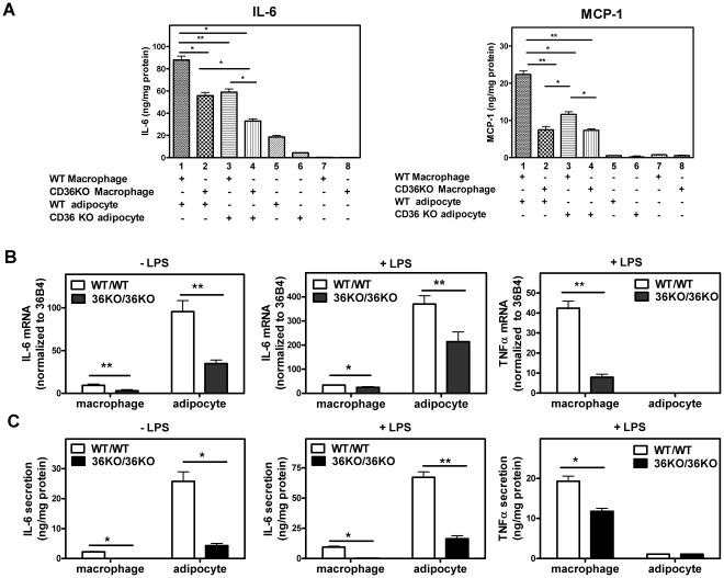
Figure 6. Macrophages and adipocytes display a synergistic and CD36- dependent cytokine response to LPS. A.
. Contact co-culture. Adipocytes were differentiated from the SVF of WT and CD36 KO mice as described in Materials and Methods. Peritoneal macrophages isolated from WT and CD36 KO mice were then layered and cultured on top of the differentiated adipocytes and co-cultured for 16 h. LPS (10 ng/mL) was then applied to the co-cultures for 4 h after which medium and cells were collected for cytokine determination. B, C. Non-contact co-culture in transwells. Primary pre-adipocytes were seeded in the bottom chamber and differentiated into mature adipocytes. Primary peritoneal macrophages were then seeded in the transwell inserts and cells were then co-cultured for 16 h. For LPS treatment groups, both adipocytes and macrophages were exposed to LPS (10 ng/mL) for 4 h after which cellular gene expression was measured by Q-PCR (B). Medium was collected and secreted cytokines were measured by ELISA (C). Values shown are mean ± SD (n = 4). *, p<0.05, **, p<0.001.

