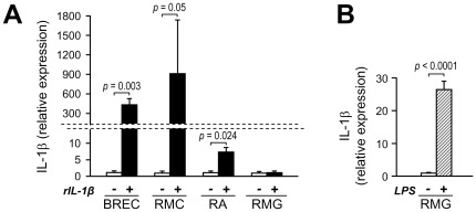
Figure 4. IL-1β autostimulation in retinal endothelial, macroglial, and microglial cells.
(A) Effect of IL-1β on IL-1β expression. Confluent culture of BREC, RMC, RA, and RMG were pre-incubated in serum-free medium for 24 hours before stimulation with rIL-1β (bovine rIL-1β for BREC; human rIL-1β for RMC, RA and RMG) at 10 ng/ml of medium. Cells were harvested after 4 hours and IL-1β mRNA levels were assayed by quantitative RealTime RT-PCR. Bars represent mean ± SD of the results obtained from different isolates of each cell type. BREC, RMC, and RA: n = 3, RMG: n = 4. (B) Effect of LPS on IL-1β expression in microglia. RMG confluent cultures were pre-incubated in serum-free medium for 24 hours before stimulation with LPS at 1 µg/ml of medium for 4 hours. The expression of IL-1β was determined by RealTime RT-PCR. Bars represent mean ± SD of the results obtained from two isolates.

