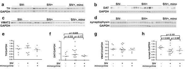
Fig. 3.
Western blot analysis indicated no overt loss of dopaminergic projections to the basal ganglia in SIV-infection. a, e Quantitation of TH in the basal ganglia by Western blot analysis showed similar levels of TH among uninfected, SIV-infected, untreated and SIV-infected, minocycline-treated macaques. b, f Measurement of DAT in basal ganglia showed no significant difference between uninfected and SIV-infected, untreated macaques. c, g VMAT2 was unchanged in SIV infection, regardless of treatment. d, h Synaptophysin was moderately reduced in SIV infection, but was not rescued by minocycline treatment. Proteins of interest were normalized to GAPDH. Bars represent medians. Statistical comparisons were made using the Mann–Whitney test

