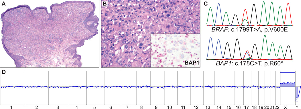
FIGURE 6.
AST from the upper back of a 44-year-old woman (case 6). A, Small polypoid melanocytic tumor (B) with medium-to-large epithelioid cells and admixed lymphocytes. Inset: Absent BAP1 staining in melanocytes but strong nuclear staining in admixed lymphocytes. C, Sequencing electropherograms show a BRAFV600E mutation and an inactivating, nonsense mutation of BAP1 (c.178C > T, p.R60*). D, Array CGH shows a balanced profile with no gains and losses.

