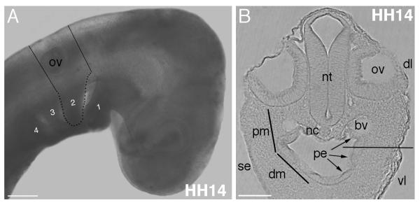
Figure 1. Location of tissues and anatomical landmarks for the explant cultures.
(A) Lines on HH14 embryo show the level of cuts to remove a transverse slice of the head and 2nd arch region. Pharyngeal arches numbered 1-4, with 2nd arch outlined in black dots. (B) A section through the level of the otic vesicle and 2nd arch region showing the tissues present. Proximal to distal mesenchyme continuum indicated by lines on the left side. Pharyngeal endoderm lines the oral cavity (arrowed). Horizontal line through arch on right side indicates approximate level of cut to exclude ventral arch mesenchyme from the explants. Abbreviations: bv, blood vessel; dl, dorsolateral level; dm, distal mesenchyme; nc, notochord; nt, neural tube; ov, otic vesicle; pe, pharyngeal endoderm; pm, proximal mesenchyme; se, surface ectoderm; vl, ventrolateral level. Scale bars: A, 200 μm, B, 100 μm.

