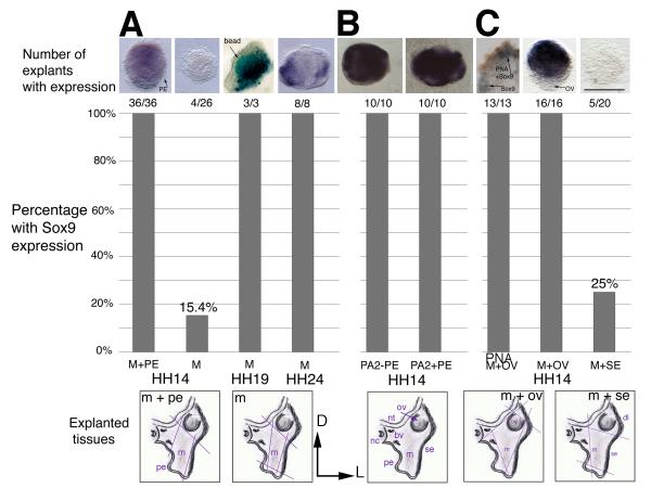
Figure 2. Sox9 expression in explants from the proximal 2nd arch region.
The schematics below the graph illustrate the tissues in the region. Orientation is dorsal to the top and lateral to the right. The colored lines indicate the cuts made to extirpate the tissue. The graph shows the number and percentage of explants that tested positive for Sox9. The tissues included in each graph are listed on the horizontal axis. (A) Explants were removed from chick embryos at HH14, HH19 or HH24 and grown in collagen gel culture to E6. Mesenchyme cultured with pharyngeal endoderm from HH14 tissues expresses Sox9 at E6, whereas mesenchyme alone does not. At HH19 and HH24, Sox9 is expressed in the mesenchyme at E6 in the absence of pharyngeal endoderm. (B) Transverse sections of the head at the level of the 2nd arch with or without pharyngeal endoderm present express Sox9 in all cases. The schematic below is labeled with the tissues found in this region at the time of culture. (C) Extirpation of the otic vesicle with mesenchyme results in PNA and Sox9 expression. Mesenchyme explanted with surface ectoderm expresses Sox9 in a quarter of cases. Abbreviations: bv, blood vessel; D, dorsal; dl, dorsolateral; L, lateral; m/M, mesenchyme; nc, notochord; nt, neural tube; ov/OV, otic vesicle; PA2, 2nd pharyngeal arch level tissue; pe/PE, pharyngeal endoderm; PNA, Peanut Agglutinin Lectin; se/SE, surface ectoderm. Scale bar: 100 μm.

