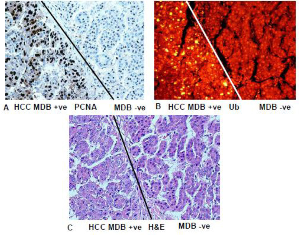
Fig 11.
Liver hepatocellular carcinoma has formed MDBs to the left of the line and not to the right of line in A, B, and C. A) PCNA stain shows that most of the nuclei in tumor cells that have formed MDBs stain positive. B) MDBs formed by the tumor cells stain positive for ubiquitin (yellow). C) Hematoxylin and eosin stain. The tumor cells that formed MDBs have vesicular nuclei compared to the tumor cells that have not formed MDBs on the right. (A×346), (B×346), (C×346).

