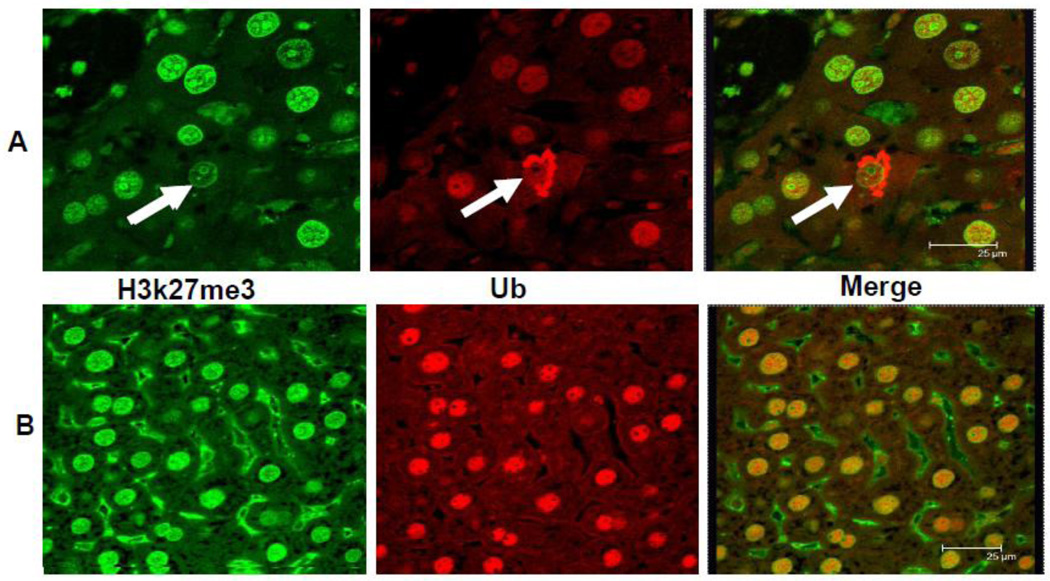
Fig 3.
The Mallory-Denk body (MDB) hepatocytic balloon cell is shown in the upper panel. The stain for H3K27me3 in the nucleus of the balloon cell is less intense than the adjacent normal hepatocytic nucleus (arrow) and the nuclei in the control liver in the lower panel. The balloon cell MDB stains positive for ubiquitin (upper panel). The merged photo of the balloon cell further documents the reduced nuclear staining for H3/K27me3 (Confocal microscope).

