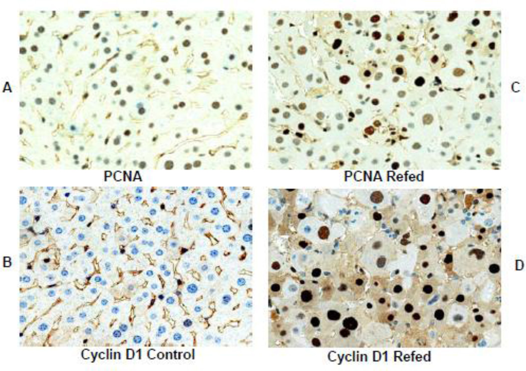
Fig 6.
Sections of liver from mice refed DDC and control stained with an antibody to PCNA showing rare nuclear staining positive in the control (A) and numerous positive staining in the DDC refed mouse liver (C). Similarly liver from the mouse refed DDC stained for cyclin D1 (D) shows numerous positive stained nuclei. The control mouse liver shows only a few positive stained nuclei. Magnifications are; (A×606), (B×606), (C×606), (D×910).

