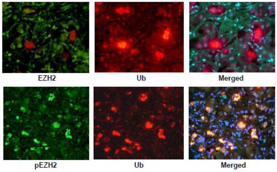
Fig 7.
A liver biopsy from a patient who had alcoholic hepatitis was stained with an antibody to EZH2 (upper panel) and an antibody to pEZH2 (lower panel). Note that the nuclei stained for EZH2 were all stained with the same intensity (left photo). The MDBs were stained green for pEZH2, red for ubiquitin and yellow when the tricolor filter was used indicating that pEZH2 was increased in MDBs. pEZH2 was also increased in the Western blot in mice refed DDC (Fig 1) and the liver section stained with the pEZH2 antibody (Fig 2). (Upper and Lower Panel×436).

