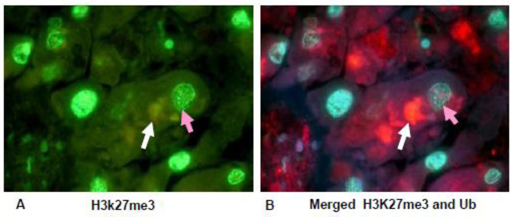
Fig 9.
Liver section from a patient who died with alcoholic hepatitis, stained for H3K27me3 and ubiquitin. Note that the liver cell containing an MDB (white arrow, red) B) stained with less intensity for H3K27me3 (orange arrow) when compared to the nucleus of a neighboring hepatocyte. (A×1040), (B×1040).

