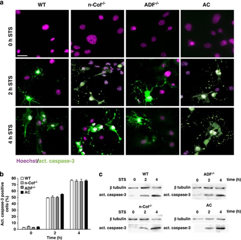
Figure 4.
STS-induced activation of caspase-3 is unchanged in the absence of ADF/cofilin activity. (a) Activation of caspase-3 was unchanged in n-Cof−/−, ADF−/−, or AC-MEFs after 2 or 4 h of STS treatment. Caspase-3 activation was analyzed by immunofluorescence using an antibody against active caspase-3 (green). MEFs were counterstained with the nuclear marker Hoechst 33342 (magenta). Scale bar corresponds to 30 μm in all panels. Representative images of six independent experiments are shown. (b) Quantification revealed similar percentages of active caspase-3-positive cells in all four MEF lines after 2 or 4 h of STS treatment. (c) Caspase-3 was activated similarly in all four MEF lines upon STS exposure. The cleaved fragment of activated caspase-3 was detected in whole-cell lysates using western blotting. Representative images of three independent experiments are shown

