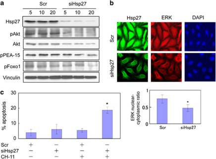
Figure 6.
Hsp27 regulates phosphorylation status of Akt and PEA-15, nuclear translocation of ERK and Fas-induced apoptosis in androgen-independent PC-3 cells. (a) PC-3 cells were transfected with various doses (5–20 nM) of Scr or siHsp27. Forty-eight hours after transfection, immunoblotting was performed to analyze the total amounts and phosphorylation states of the indicated proteins. (b) Nuclear translocation of ERK was assessed by immunofluorescence microscopy after transfection of PC-3 cells with 20 nM Scr or 20 nM siHsp27. Cells were fixed 18 h after culturing in low-serum (0.5%) conditions and were assessed by immunofluorescence staining with anti-Hsp27 (green) and anti-ERK (red) antibodies. DAPI (blue) nuclear counterstaining was used for marking cell nuclei. The mean fluorescence ratios FN/C are shown for ERK. Values shown represent mean±S.D. The symbol ‘*' denotes statistical significance (P<0.05). (c) Assessment of the effect of Hsp27 knockdown on Fas-induced apoptosis of PC-3 cells. PC-3 cells were transfected with 20 nM Scr or 20 nM siHsp27. At 48 h after transfection, apoptosis in response to the agonistic anti-Fas antibody, CH-11, was assessed by flow cytometric analyses of the proportion of cells containing subdiploid DNA as described above

