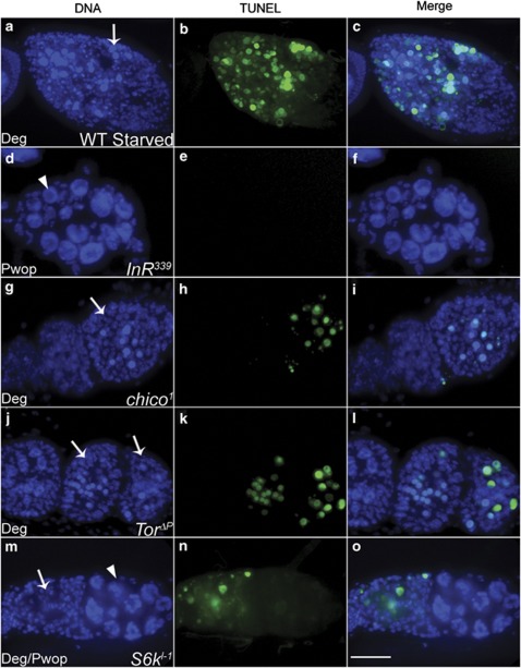
Figure 4.
DNA fragmentation is blocked in insulin signaling mutant Pwop egg chambers. All egg chambers were assayed for fragmented DNA using the TUNEL ApopTag Plus Fluorescein In Situ Detection Kit (green), and stained with DAPI (blue). (a–c) WT degenerating egg chamber, in which NC nuclear fragmentation has occurred, shows many TUNEL-positive spots. (d–f) InR339 GLC stage 5 Pwop egg chamber. (g–i) chico1 GLC healthy early stage egg chambers and a degenerating stage 2 egg chamber. (j–l) TorΔP GLC healthy (stage 4) and degenerating (stages 4 and 5) egg chambers. (m–o) S6kl−1 GLC degenerating (stage 1) and Pwop stage 4 egg chambers. Arrows indicate degenerating egg chambers and arrowheads indicate Pwops. All images were taken at the same magnification and are projections of 15–20 images. Scale bar is 15 μm

