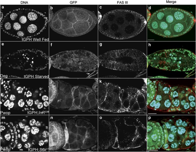
Figure 6.
PI3K activity is lost in InR and S6k Pwops. All egg chambers expressing the tGPH reporter (green) were stained with DAPI (blue) and Fas III (red) to label FCs. FasIII is enriched in the polar cells in mid-stage egg chambers. (a) tGPH mid-stage egg chamber from well-fed fly displayed (b) PI3K activity at the plasma membranes of FCs and NCs. (c) Fas III and (d) Merge indicates co-localization at the FC plasma membrane of tGPH reporter and Fas III. (e) tGPH-degenerating mid-stage egg chamber from starved fly displayed (f) no PI3K activity at the plasma membranes of the FCs or NCs. (g) Fas III staining shows that FCs increased in size during engulfment of NC remnants. (h) Merge. (i–k) tGPH;InR339 GLC Pwop stage 4 egg chambers. (l) Merge indicates co-localization at the FC plasma membrane of tGPH reporter and Fas III. (m–o) tGPH;S6kl−1 GLC Pwop stage 3 egg chamber. (p) The merge displays co-localization at the FC plasma membrane of tGPH reporter and Fas III. Scale bar is 20 μm

