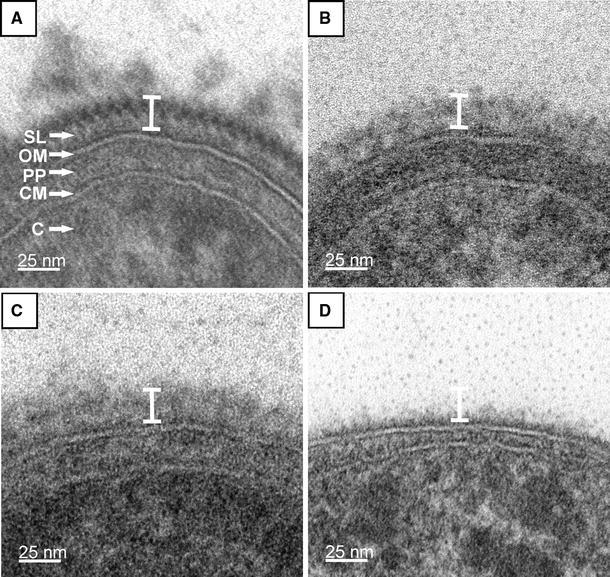
Fig. 3.

Ultra-thin cross-sections of whole cell preparations from T. forsythia. a T. forsythia wt cells with a 2D crystalline S-layer; b T. forsythia ΔtfsB mutant cell; c T. forsythia ΔtfsA mutant cell. In either S-layer single mutant the S-layer is not visible as a cross-sectioned 2D-crystal, but an unstructured “stained mass” without periodicity is present on top of the outer membrane, which is less densely packed than the S-layer on wild-type cells. d T. forsythia ΔtfsAB double mutant without an S-layer. The white bar indicates that the overall thickness of the S-layer is identical in T. forsythia wt and T. forsythia S-layer single-mutant cells. SL S-layer, OM outer membrane, PP periplasmic space, CM cytoplasmic membrane, C cytoplasm
