
Fig. 1.
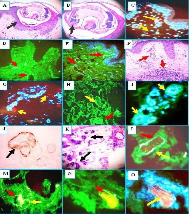
H & E (a, b, f, k): see Results section for description. DIF (c, d, e, g,h ,i, l, m, n, o). Note that nuclei are counterstained with 4’, 6-diamidino-2-phenylindole (Dapi) on (c, e, g, and o) (blue stain). c. We found some overexpression of ezrin in the epidermal area close where the mite was found in the epidermis (red dot staining) (yellow arrows). d. and e. Positive staining of the superficial and intermediate vessels with antibody to human fluorescein isothiocyanate (FITC) conjugated fibrinogen (green staining) (red arrows).g we found some overexpression of junctional adhesion molecule (JAM-1) around the inflamed vessels close to the scabies infection site (red dot staining) (yellow arrows). h, i, and l, Note positive staining to the eccrine sweat glands utilizing anti-human FITC conjugated IgG antibody (green staining) (yellow arrows). j Positive immunohistochemistry (IHC) staining to the sweat glands using Complement/C5b-99/MAC) (brown staining) (black arrow). m, n Note autoreactivity to a nerve utilizing the FITC conjugated human albumin antibody (green stain) yellow arrow that colocalize with ppg.9.5 neural marker (yellowish staining) (red arrows. In n, higher magnification. In o, we present the same area as in m and n, but we utilize a Texas red conjugated antibody to human glial fibrillary acidic protein and a FITC conjugated human fibrinogen antibody to confirm the neural nature of this structure. The overlapping of red and green stain shows like a bright pinkish (yellow arrow). The nerve was also located in proximity to the mite.