Figure 2.

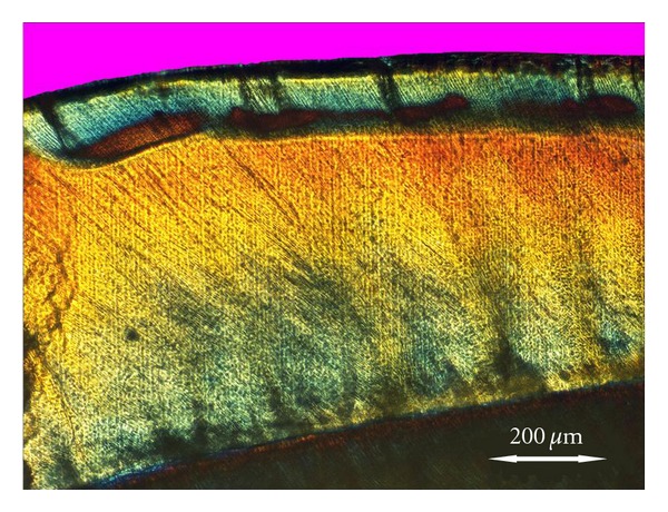
POLMI image, at 4x. The lesion is seen as a uniform demineralized zone, with a positive birefringent bulk below a negatively birefringent surface layer. At the lesion front, a zone with a higher degree of demineralization is seen.

POLMI image, at 4x. The lesion is seen as a uniform demineralized zone, with a positive birefringent bulk below a negatively birefringent surface layer. At the lesion front, a zone with a higher degree of demineralization is seen.