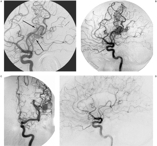
Figure 2.
Rolandic BAVM, previously embolized at another institution with occlusion of the Rolandic artery. The BAVM was supplied by an extensive pial arterial collateral system. A) Angiography with injection into the left internal carotid artery. Lateral view in the early phase of contrast media passage, just as the vein becomes visible. Two distinct shunting zones are evident (arrows) which were separately delineated before GKRS. B) Later phase of the angiogram, when the vein is filled with contrast medium. It is no longer possible to distinguish the shunts. The two separate target delineations are shown on this image. C) Posteroanterior view in a late phase of the angiogram with the two separate target delineations. Treatment was with 25Gy to the 50% isodose. D) After two years the BAVM was obliterated. There is no remaining arteriovenous shunt. Note that the pial arterial collateral network has regressed almost totally.

