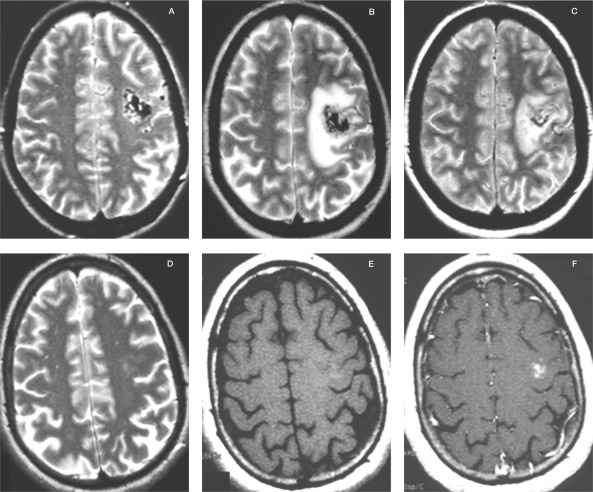
Figure 5.
MRI of a left frontal BAVM treated with a dose of 25Gy to the periphery in the GK. A) T2-weighted image at the time of the treatment. B) T2-weighted image eight months after the treatment. The patient developed seizures. C) T2-weighted image 12 months after the treatment. The patient was on steroids and anticonvulsive medication. D) T2-weighted image 24 months after the treatment. The patient was no longer on steroids or anticonvulsive drugs. E) T1-weighted image without Gadolinium 24 months after the treatment. F) T1-weighted image with Gadolinium 24 months after the treatment. Note the contrast enhancement at the position of the previously treated BAVM. The malformation was proven by angiography to be obliterated.

