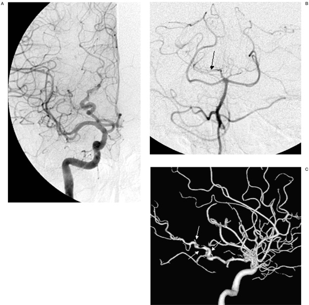
Figure 2.
Selective injections of the right internal carotid artery (A) and right vertebral artery (B) and 3D DSA shaded surface display (SSD) reconstruction (C) demonstrate a series of alternating dilatation and segmental narrowing of the P3 segment of the right PCA (pearl and string sign, white arrows). The right vertebral artery injection (B) shows washout of the contrast media (arrow) in the right PCA due to un-opacified bloods from right posterior communicating artery.

