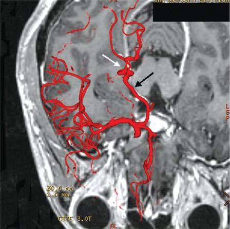
Figure 3.
Axial fusion image clearly showing the normal P2 segment of the right PCA located between the cerebral peduncle and the tentorium. The abnormal P3 segment (white arrow) is seen to cross the free edge twice from medial to lateral proximally and lateral to medial more distally. Note that there is nearly perfect registration of the fused DSA image to the corresponding enhanced right PCA in the MRI (black arrow).

