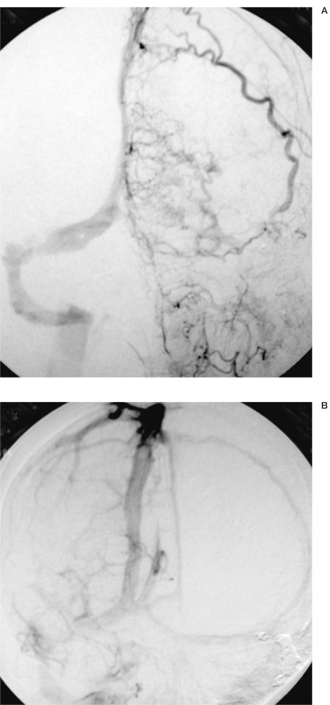
Figure 4.
A:) A left external carotid angiogram showing persistent disappearance of the previously treated dural AVF at the left sigmoid sinus and a newly developed dural AVF at the superior sagittal sinus supplied by the left superficial temporal artery. B) A right internal carotid angiogram (venous phase), showing left transverse sinus and superior sagittal sinus.

