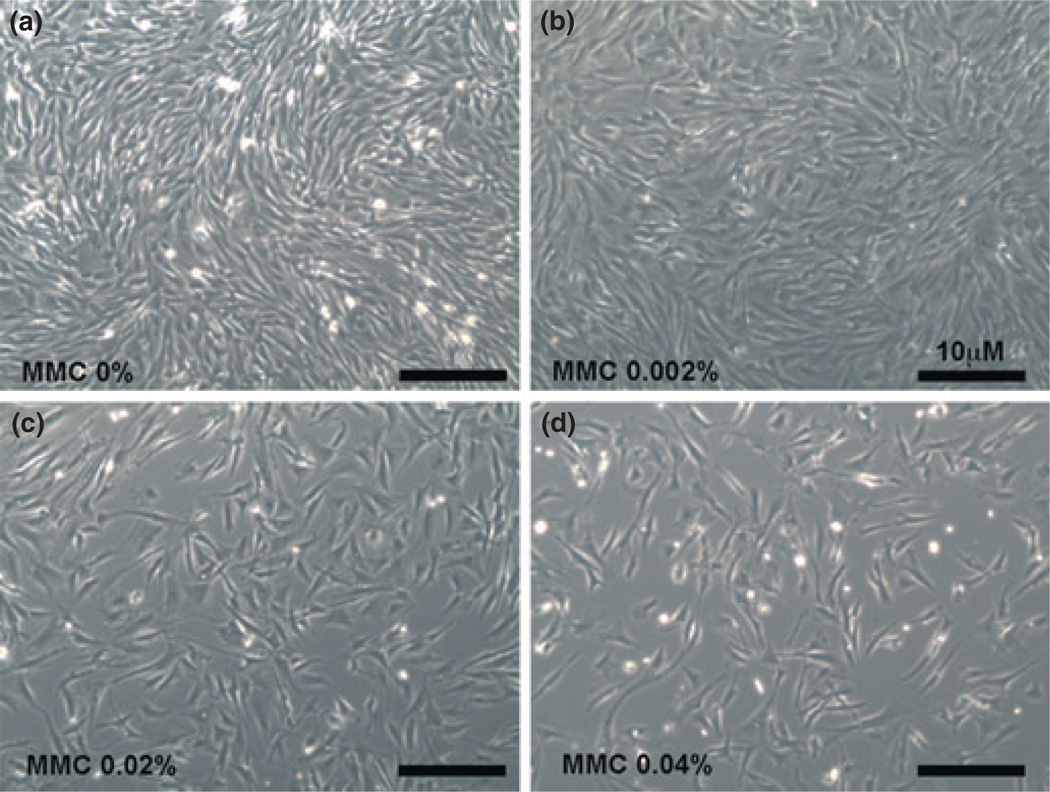
Figure 1.
Dose-dependent treatment of mitomycin C (MMC) in canine stromal fibroblast (CSF) primary cultures. Bright-field images of CSF cultures were taken using a light microscope. (a) Untreated control and (b) 0.002% MMC demonstrated no change in cellular morphology. A single 2 min dose of 0.02% MMC (c) did not alter cellular morphology. Higher doses of MMC (0.04%, d) reduced CSF populations considerably without altering phenotype or viability. The anti-fibrotic effects of MMC at low dose and short exposure times are likely due to proliferation or growth inhibition.

