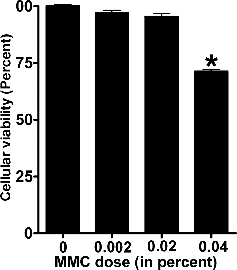
Figure 2.
Percent viability of canine stromal fibroblasts (CSFs) treated with increasing doses of mitomycin C (MMC). CSFs treated with various concentrations of MMC were stained with trypan blue reagent to count the number of viable cells. MMC dose of 0.002% and 0.02% only slightly decreased cellular viability but were not statistically significant. However, 0.04% significantly reduced the cellular viability up to 27% (P < 0.001). Error bars indicate standard error.

