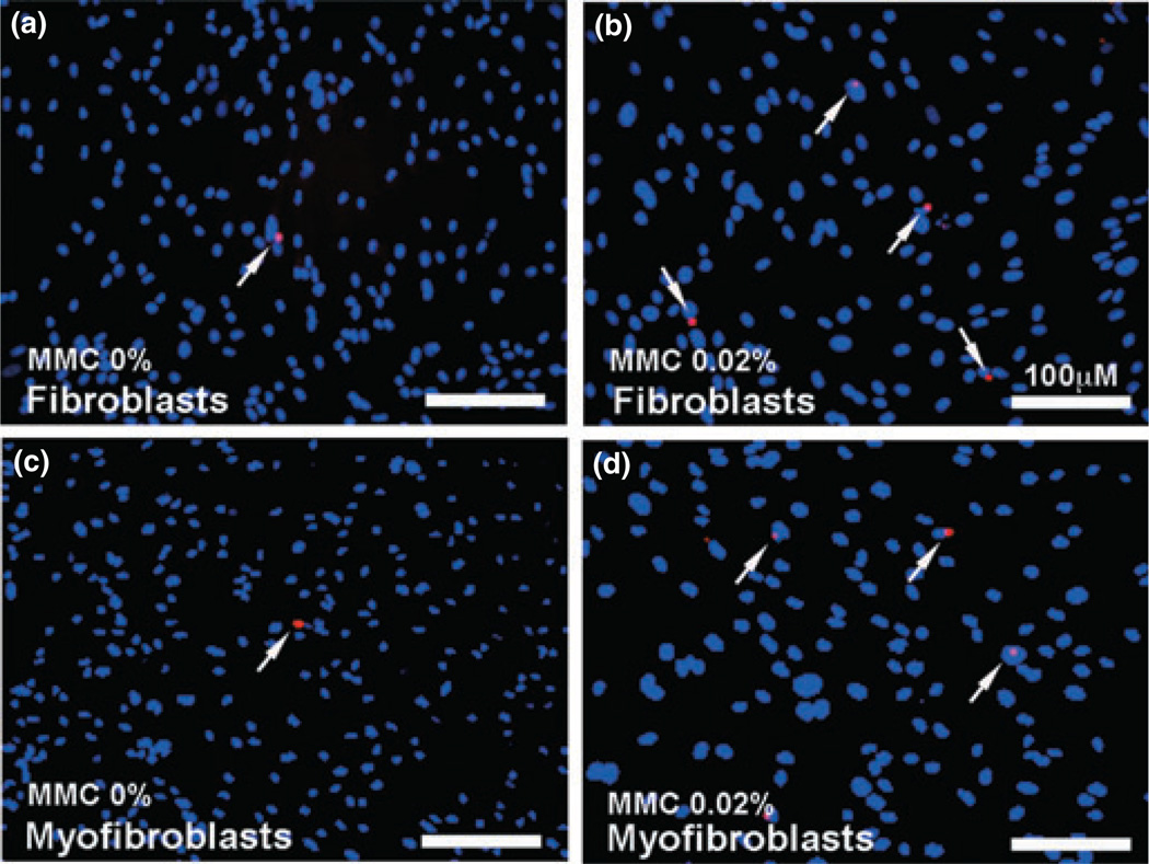
Figure 3.
Terminal deoxynucleotidyl transferase dUTP nick end labeling (TUNEL) assay demonstrating apoptotic cells in fibroblast and myofibroblast cultures treated with mitomycin C (MMC). Canine corneal fibroblasts (a, b) as well as TGFβ-induced myofibroblasts (c, d) were exposed to 0.02% MMC for 2 min. Based on six nonover-lapping fields quantification, MMC untreated controls (a, c) demonstrated 1–4 TUNEL-positive cells (red) whereas MMC-treated cultures (b, d) showed 3–8 TUNEL-positive red cells. The 0.02% MMC treatment for 2 min to canine corneal fibroblast and myofibroblast did not induce significant cell death. Arrows indicate apoptotic cells. Nuclei are stained blue with DAPI; scale bar denotes 100 µm.

