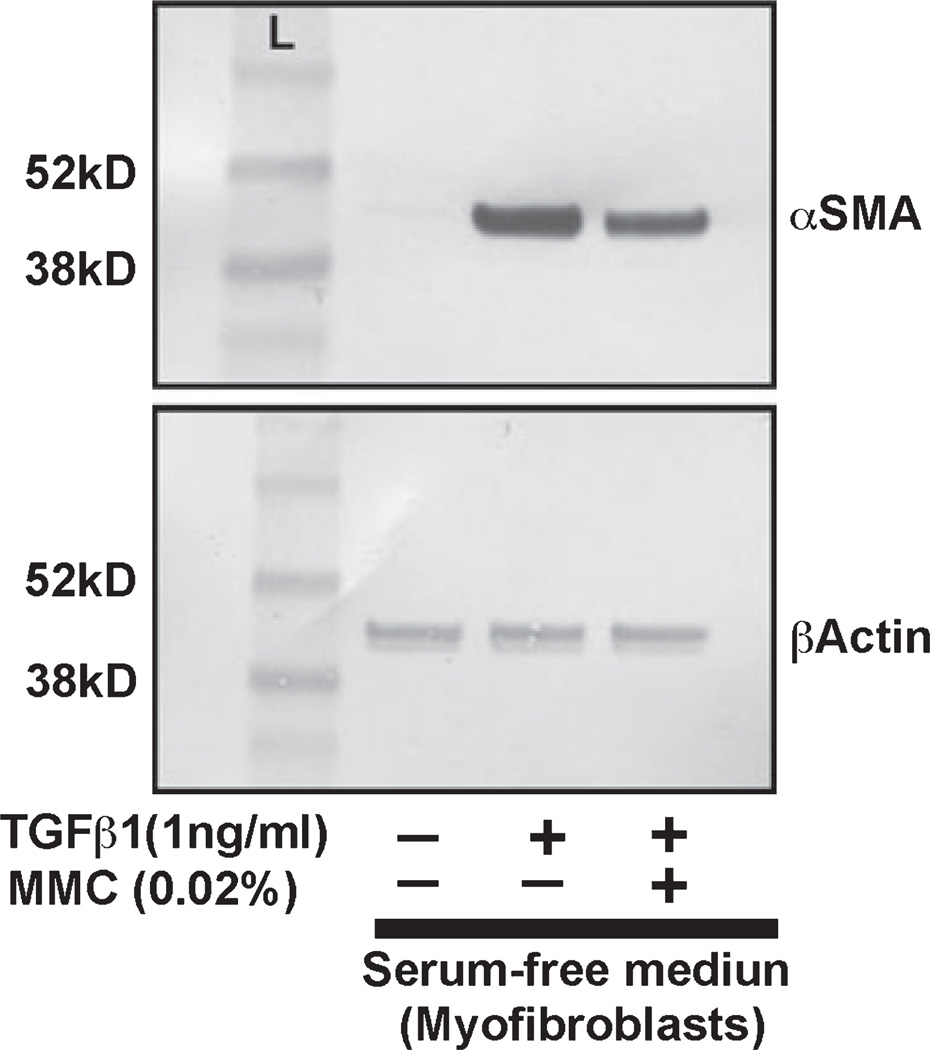
Figure 7.
Immunoblot analysis demonstrating quantitative measurement of alpha smooth muscle actin (αSMA) (myofibroblast marker) in canine stromal fibroblast (CSF) cultures treated with or without TGFβ1 and 0.02% mitomycin C (MMC). Equal quantity of protein (20 µg) was loaded in each lane. Lane 1 is the untreated control. Lysates prepared form TGFβ-treated cells were loaded in lane 2. TGFβ + MMC treated cell lysates were loaded in lane 3. Beta-actin was used as house keeping gene. A single MMC treatment (0.02% for 2 min) (lane 3) showed significant decrease in TGFβ1-induced myofibroblast formation (lane 2) in CSFs compared to untreated controls (lane 1).

