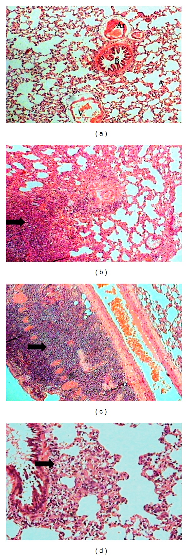
Figure 4.

(a) Lung section of control group with normal histological appearance of alveolus (A), bronchiole (B), arteriole (Ar) and venule (V) (HE, 10X). (b) Lung of rats with induced sepsis showing different degrees of lung consolidation and alveolar spaces were infiltrated with a large number of mononuclear cells (HE, 10x). (c) Presence of mononuclear infiltrated in the wall of bronchiole (HE, 10x). (d) Local thickening of infiltrated mononuclear cells in alveolar wall and peribronchial area (HE, 20x).
