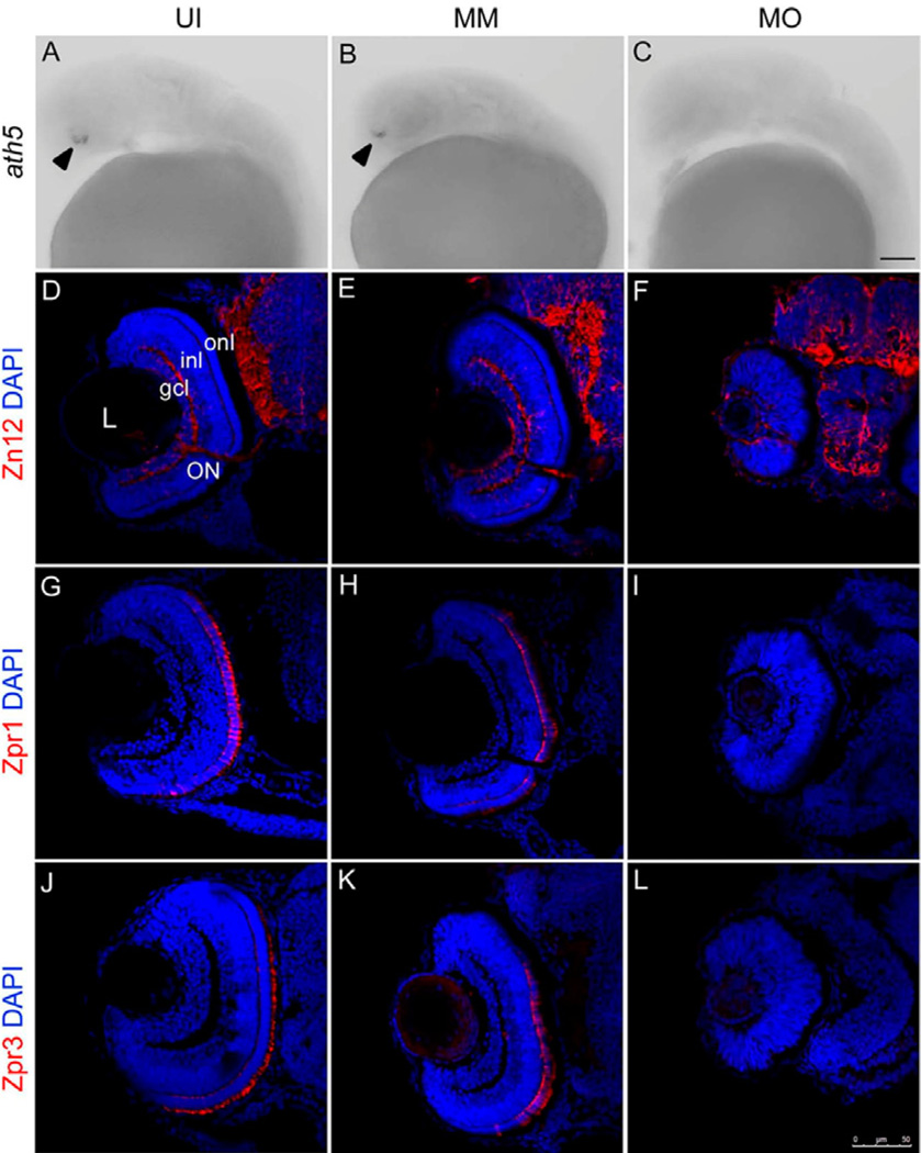
Figure 2. fms knockdown delays neurogenesis initiation at 28 hpf and affects neuronal differentiation at 72 hpf.
Panels A–C show the in situ analysis of ath5 expression during retina development in the retina of uninjected (UI), mismatch control (MM) and fms morphants (MO) at 28 hpf. Note that the expression of ath5 was detected in the retina of uninjected and mismatch controls (arrowheads), but was absent in fms morphant retina. Panels D–L illustrate sections taken through the retinas at 72 hpf. Panels D–F illustrate Zn12 staining; panels G–I illustrate Zpr1 staining; panels J–K illustrate Zpr3 staining. Note that in the fms morphant retinas, only a handful of differentiated Zn12-positive cells were present at 72 hpf, possessed rudimentary optic nerves and had the appearance of an undifferentiated neuroepithelium. Neither Zpr1 nor Zpr3 was detected, while the retinas are well laminated and differentiated, showing strong expression of Zn12, Zpr1 and Zpr3 in the uninjected and mismatch control larvae at 72 hpf. L, lens; gcl, ganglion cell layer; inl, inner nuclear layer; onl, outer nuclear layer; ON, optic nerve. Scale bar: A–C, 50µm; D–L, 75 µm.

