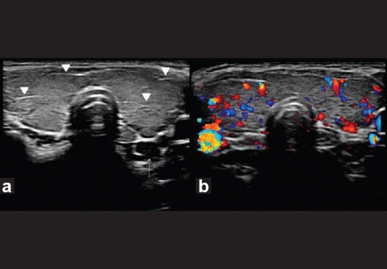
Figure 5.

Hashimoto's thyroiditis. Transverse gray-scale ultrasound (a) and color Doppler (b) neck, of a 35-year-old female patient, who presented with features of hypothyroidism and had antithyroid antibodies positive for the disease, demonstrates diffuse enlargement of thyroid gland with linear echogenic fibrous bands (arrowheads) but normal vascularity. Note a small hypoechoic lymph node (arrow) in posterior aspect of inferior pole of left lobe of the thyroid gland
