
Figure 6.
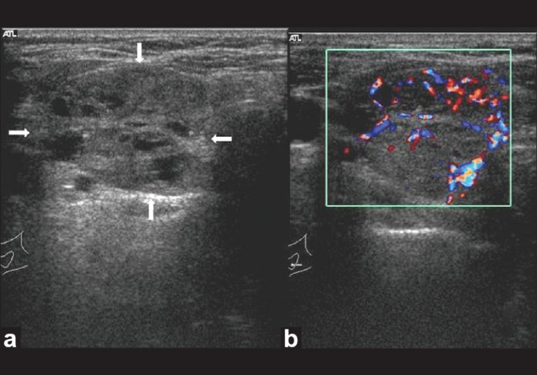
Follicular lesion thyroid. Transverse gray-scale ultrasound (a) and color Doppler (b) neck, of a 40-year-old female patient, shows a large well circumscribed iso-hypoechoic solid thyroid nodule with multiple internal cystic spaces (arrow) and both central and peripheral vascularity