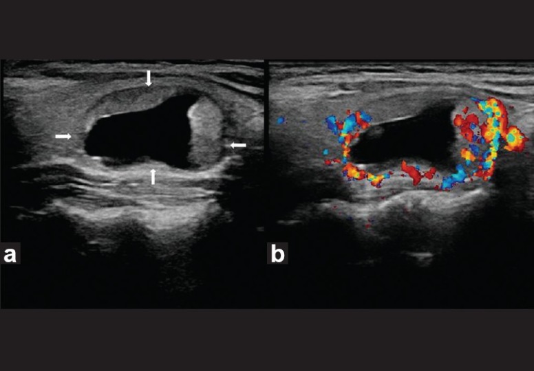
Figure 7.

Benign thyroid nodule with large intratumoral cyst. Transverse gray-scale ultrasound (a) and color Doppler (b) neck, of a 55-year-old female patient, reveal a well-circumscribed right-sided thyroid nodule (arrow) with a large intratumoral cyst and solid peripheral component which shows increased vascularity. The lesion demonstrates a thin hypoechoic rim and posterior acoustic enhancement
