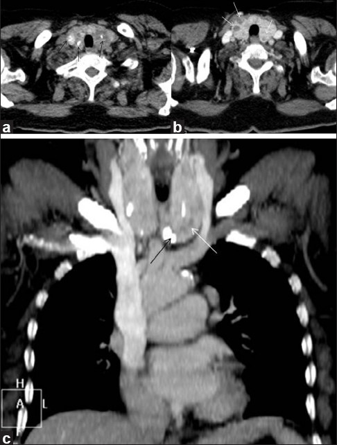
Figure 9.

Multinodular goiter. Plain axial (a) and contrast-enhanced axial (b) and coronal (c) CT scan neck region, of a 52-year-old female patient, shows enlargement of bilateral thyroid lobes due to presence of multiple non-enhancing hypodense (thin white arrows) and calcified thyroid nodules (thin black arrows)
