
Figure 1.
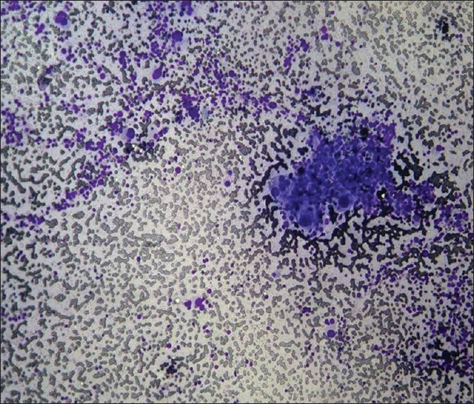
Photomicrograph of fine needle aspiration from the right adrenal mass, showing sheets of dispersed cells with foamy fragile cytoplasm, uniform, enlarged and hyperchromatic nuclei with inclusions and multi-lobed nucleoli
Official websites use .gov
A
.gov website belongs to an official
government organization in the United States.
Secure .gov websites use HTTPS
A lock (
) or https:// means you've safely
connected to the .gov website. Share sensitive
information only on official, secure websites.
Photomicrograph of fine needle aspiration from the right adrenal mass, showing sheets of dispersed cells with foamy fragile cytoplasm, uniform, enlarged and hyperchromatic nuclei with inclusions and multi-lobed nucleoli