Figure 3.

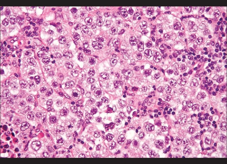
Histopathology of the biopsy specimen - large, round cells with vesicular nuclei and clear or finely granular cytoplasm that is eosinophilic admixed with lymphocytes in the stroma, confirming the diagnosis of dysgerminoma (H and E, ×300)

Histopathology of the biopsy specimen - large, round cells with vesicular nuclei and clear or finely granular cytoplasm that is eosinophilic admixed with lymphocytes in the stroma, confirming the diagnosis of dysgerminoma (H and E, ×300)