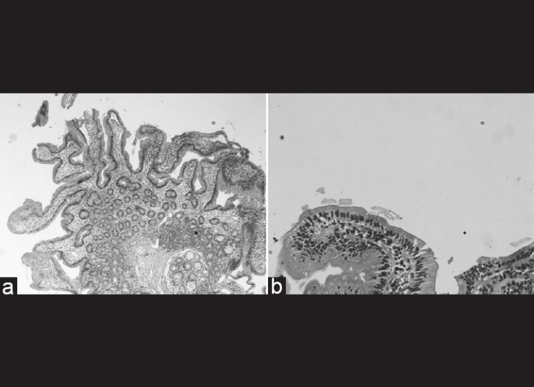
Figure 1.

(a) Giemsa stained duodenal biopsy (×4) shows villus to crypt ratio of 3:1,intraepithelial lymphocytes are not increased,no luminal parasite identifed,there is mild chronic inlammatory cell infiltrate in the lamina propria features are non specific. (b) Giemsa stained duodenal biopsy (×20) - one villus with same features as desribed
